The human heart is fundamentally a fist-sized muscular organ located in the thoracic (chest) cavity. Small as it is, the heart plays a huge role in the functioning of the body by pumping blood around to supply organs and tissues with oxygen and nutrients, and to the lungs for purification.
Circulation
Arteries transport blood away from the heart, while veins bring blood back to the heart. Hence, all arteries carry oxygenated blood, except for the pulmonary artery which carries de-oxygenated blood to the lungs for purification. Similarly, all veins carry de-oxygenated blood, except for the pulmonary vein which carries purified, oxygenated blood back to the heart from the lungs. This oxygenated blood, which also receives nutrients from our diet along the way, is then pumped out through the aorta, the largest artery, to all the other arteries that end up as tiny blood capillaries at their respective organ or tissue sites. De-oxygenated blood from around the body is transported back to the heart through veins that eventually drain into the superior and inferior arms of the vena cava through which blood re-enters the heart.
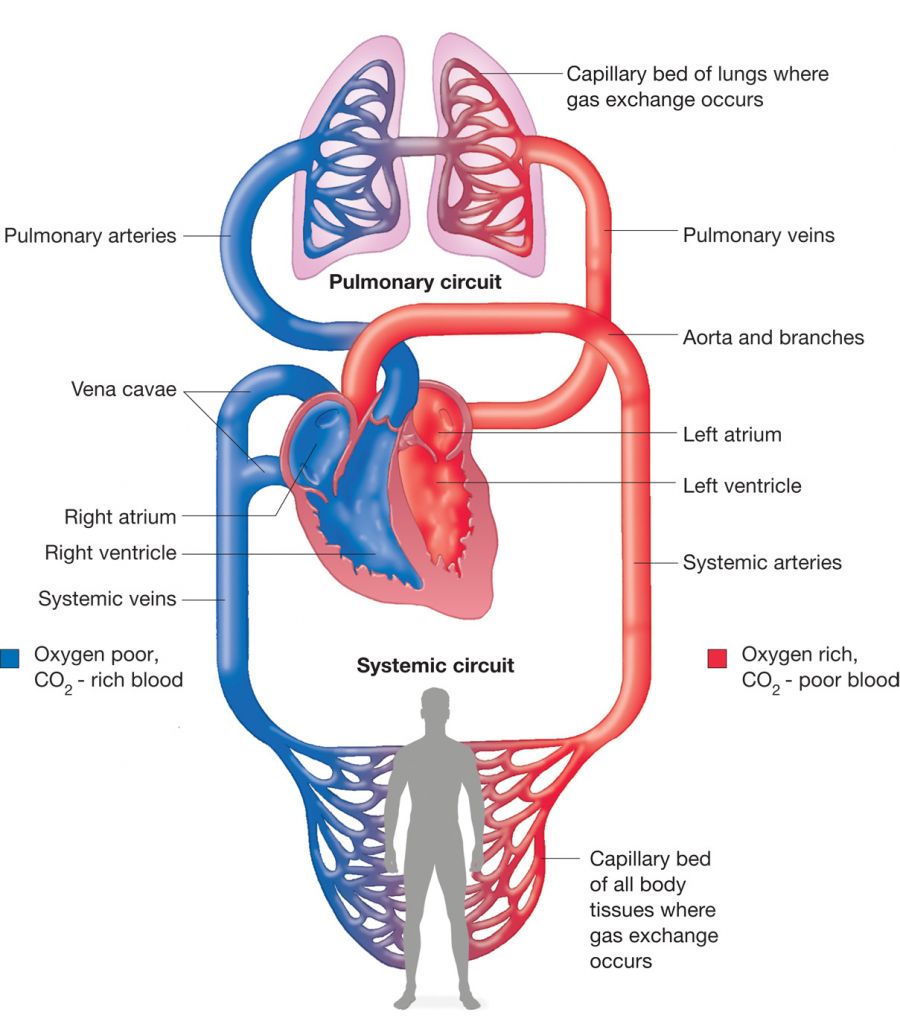
Illustration from Label Design Ideas 2020
Anatomy of the Heart
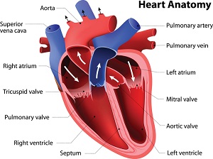
Illustration from National Institute on Aging
The heart is divided into four chambers – the right and left atria (sing. atrium) at the top, and the right and left ventricles at the bottom. The entry and exit of blood from the ventricles are controlled by the opening and closing of several valves. De-oxygenated blood from the vena cava enters the right atrium from where it is pushed down into the right ventricle by the opening of the tricuspid valve. Opening of the pulmonary valve then facilitates the pumping of this blood to the lungs via the pulmonary artery. Similarly, the opening of the mitral (bicuspid) valve allows the passage of oxygenated blood (coming from the lungs) from the left atrium into the left ventricle. The subsequent opening of the aortic valve allows this blood to be pumped out into the aorta. The tricuspid and mitral valves are collectively referred to as atrioventricular valves, while the aortic and pulmonary valves are collectively referred to as semilunar valves.
The following animation by National Geographic gives us a good idea of how this process takes place.
Conducting System of the Heart
Pumping blood from the atria to the ventricles, and then out from the heart involves a coordinated sequence of events that include the opening and closing of the above-mentioned valves, as well as contraction of the heart’s muscular walls. These activities are controlled by a series of electrical impulses, which is what we read in an electrocardiogram, or ECG. Illustrated below is an ideal voltage-versus-time graph for the electrical activity in a single heartbeat. This involves three main components: P wave (depolarisation of atria), QRS complex (depolarisation of ventricles), T wave (repolarisation of ventricles).
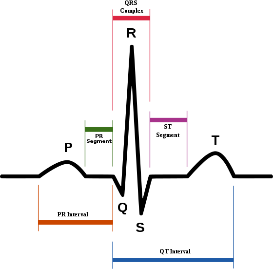
Illustration from Wikipedia
This electrical activity and subsequent contraction of heart muscles occur as a result of the coordinated effort of the heart’s nervous system. Electrical impulses generated by the sinoatrial (SA) node, also known as the heart’s pacemaker, cause atrial contraction. The atrioventricular (AV) node then sends electrical impulses down through the left and right branches of the Bundle of His and on to the Purkinje fibres in the ventricular walls, resulting in ventricular contraction which pumps blood out of the heart.
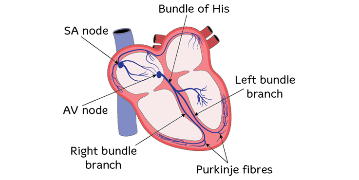
Illustration from Heart Foundation
Pressure, Pulse and the Beat
The lub-dub we hear in what we call a heartbeat is the sound produced by the closing of, firstly, the atrioventricular valves (lub) and then the semilunar valves (dub) in a single heart cycle. The entire process usually occurs within a time frame of one second or less.
With every heartbeat, blood gets pumped out of the heart and into the arteries, causing a sort of ripple that can be felt in the arteries. This is what is felt as a pulse. For a healthy adult at rest, the heart beats between 60 to 100 times every minute. Therefore, a pulse rate within the range of 60-100 per minute is considered to be normal. Thus, beating at an average rate of around 100,000 times a day and around 35 million times a year, the heart is a vital organ that needs to be taken care of by maintaining a healthy diet and active lifestyle. Pulse rate can vary with age, physical activity, excitement and illness.
When we measure blood pressure, we look at two values – systolic blood pressure and diastolic blood pressure. The higher ‘systole’ value is a measure of the pressure exerted during a heartbeat. This is the point at which blood pressure is at its highest. The lower ‘diastole’ value measures blood pressure between heartbeats, at which point it is at its lowest. The normal blood pressure for a healthy adult at rest is considered to be 120/80 mmHg, read as ‘120 over 80 millimetres of mercury’. It is very important that blood pressure is always measured when the patient is seated (or lying down), is calm and has rested for at least 20 minutes.
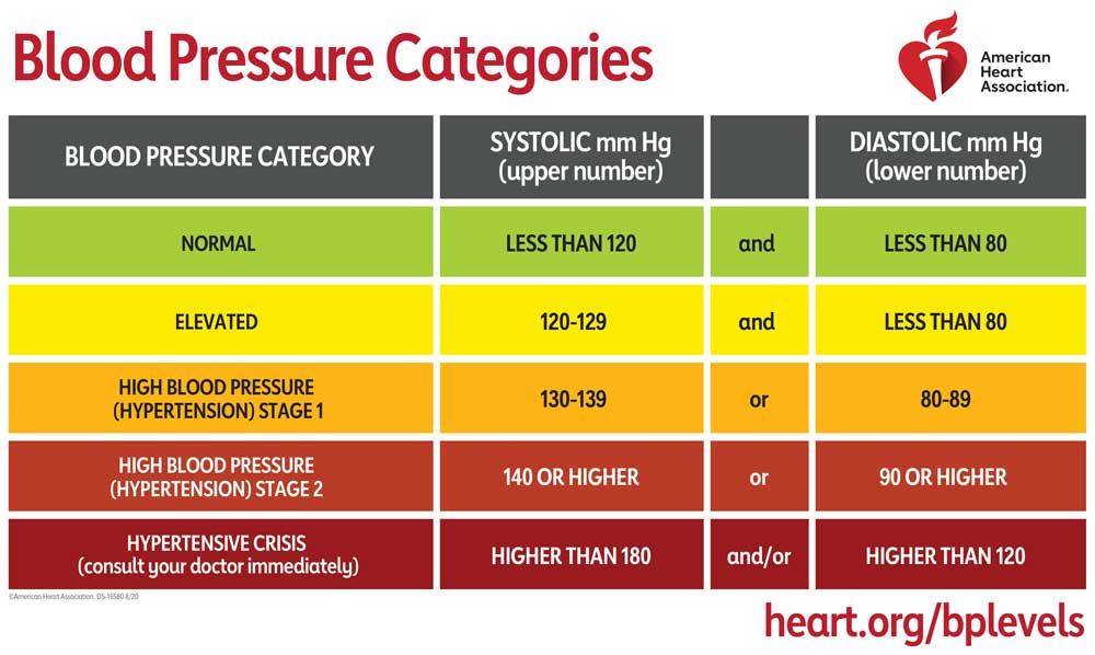
Values from American Heart Association
An increase in systolic pressure that is not a result of stress/excitement or physical activity is referred to as hypertension and could be the result of narrowed arteries (in which case the heart would have to push harder to pump blood into the arteries), among other reasons. Diastolic pressure also usually increases in such cases. Common complications that could arise from long-term hypertension include cardiac arrest (heart attack), stroke, aneurysm and heart failure.
A decrease in systolic pressure when at rest, referred to as hypotension, could result from arterial widening, dehydration, excessive bleeding or cardiac damage/weakness, among other reasons. Diastolic pressure also shows a decline in such cases. Prolonged hypotension could result in damage to the kidneys.
Changes and irregularities in blood pressure, pulse rate and heart rhythm could result from numerous ailments and medical conditions. These are therefore some of the first parameters that are measured when a patient presents themselves to a doctor. Further investigations would then be carried out in line with other observed symptoms. As such, restoring and maintaining these parameters at their normal levels is of paramount importance in any medical case.
So, what’s in a heartbeat?
The complexities involved in keeping a heart beating are many. Similarly, the service that a beating heart does to our body is tremendous. In this article, I have attempted to touch on most of these aspects in as basic and concise a manner as possible.
Simply stated, our heartbeat keeps us alive and nourished.
Cover illustration adapted from VectorStock.

3 Comments Add yours